Article Text

Abstract
A 70-year-old man was admitted to our COVID-19 ward with thoracalgia, productive mucus cough, fatigue and erythematous–violaceous macules on the inner side of feet and interdigital regions. The patient was started on oxygen and dexamethasone. On the day of discharge, he maintained the skin changes despite the resolution of COVID-19 symptoms. A 57-year-old woman initially presented with diffuse urticarial rash on the cervical and chest region. Oral cetirizine was started, and pruritus improved. Thirty days after the discharge, the patient maintained the rash, but without pruritus. A 49-year-old man was admitted with thoracalgia, shortness of breath, dry cough and urticarial rash on the cervical and chest region. The patient was treated with cetirizine. The pruritus improved, and 5 days after discharge, the urticarial areas completely disappeared.
- COVID-19
- dermatology
This is an open access article distributed in accordance with the Creative Commons Attribution Non Commercial (CC BY-NC 4.0) license, which permits others to distribute, remix, adapt, build upon this work non-commercially, and license their derivative works on different terms, provided the original work is properly cited and the use is non-commercial. See: http://creativecommons.org/licenses/by-nc/4.0/.
Statistics from Altmetric.com
Background
In December 2019, in the Chinese city of Wuhan, were reported the first cases of unexplained pneumonia,1 caused by a new infectious pathogen named SARS-CoV-2.2 The resulting disease was called COVID-193 and has become a global health threat, infecting 118 058 503 individuals, and resulting in 2 621 046 deaths by 12 March 2021.4
With the global increase in the number of SARS-CoV-2 infections, there has been a rising frequency of cutaneous manifestations with different morphology types. Mechanisms responsible for the skin rash in SARS-CoV-2 have been formulated, including direct virus infection of an open wound of the skin, immune system responses or iatrogenic drug introduction.5
In a prospective nationwide case collection study in Spain conducted by Galván et al, which included 375 patients, the authors described five clinical patterns associated with COVID-19. These patterns comprised acral areas of erythema–oedema with some vesicles or pustules (pseudochilblain), other vesicular eruptions, urticarial lesions, other maculopapules, and livedos or necrosis.6 Their frequency and timing in the disease’s natural course are still challenging to determine.7 However, some authors reported maculopapular eruptions, pseudochilblain and urticarial rash to be the most common mucocutaneous manifestations in COVID-19,8 9 developing few days before the onset of the systemic symptoms. Erythematous rash and localised or generalised urticaria appear to be the most shared cutaneous manifestations in patients with acute severe infection, although the underlying cause, such as viral infection or drug reaction may be challenging to identify.10 For example, hydroxychloroquine, experimentally used for the treatment of COVID-19 in the early days of the pandemic, is associated with a generalised pustular rash.8 10
Since cutaneous manifestations of COVID-19 seem to be self-resolving, the treatment recommendations support symptoms control with analgesics, corticosteroids or non-steroidal anti-inflammatory drugs.11
First case
Case presentation
A 70-year-old man with a medical history of arterial hypertension and type 2 diabetes mellitus was admitted to our COVID-19 ward. The diagnosis of SARS-CoV-2 infection by a positive nasopharyngeal swab test was made while he was asymptomatic, as he had high-risk contact with a family member who had been diagnosed with COVID-19. Subsequently, the patient began to experience thoracalgia, productive mucus cough and progressive fatigue and was admitted 8 days after the SARS-CoV-2 positive test to the hospital with bilateral pneumonia. He presented with erythematous–violaceous macules on the inner side of his feet (figure 1) and interdigital regions (figures 2 and 3).
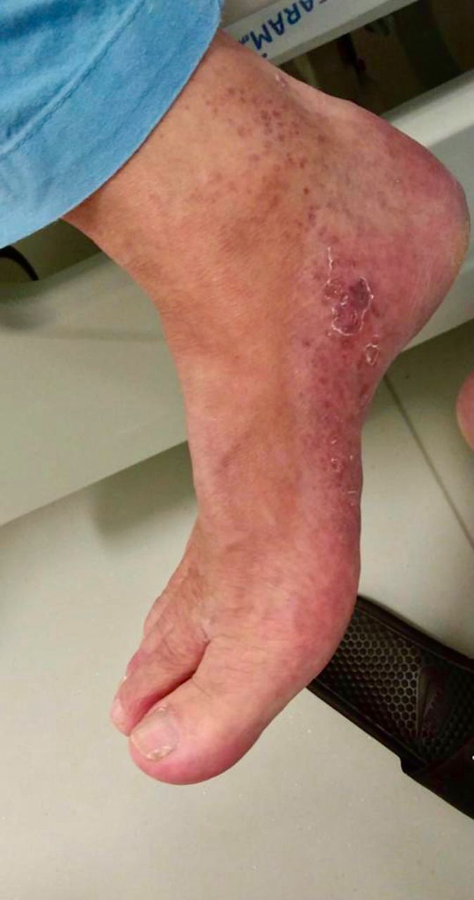
Erythematous–violaceous macules on the inner side of the right foot.
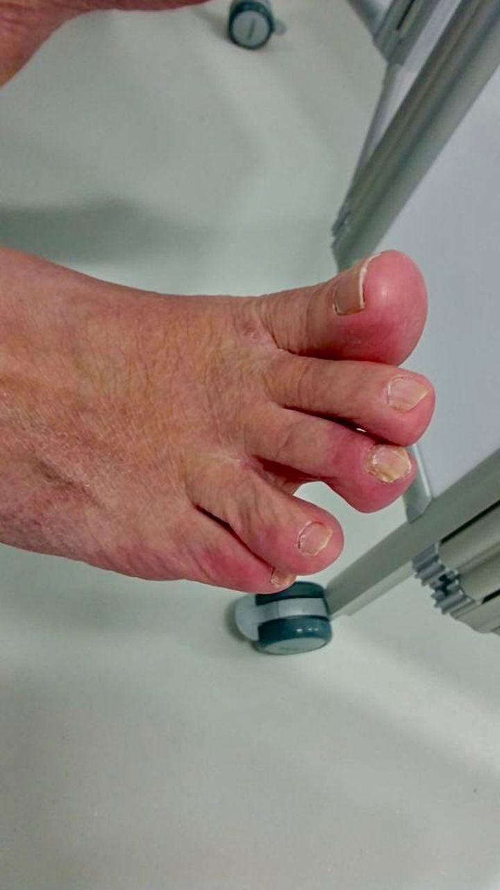
Erythematous–violaceous macules in the interdigital region.
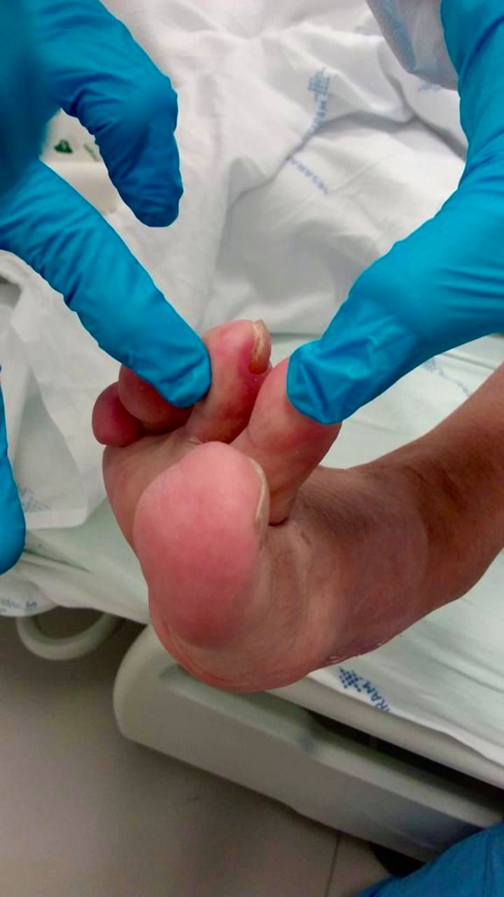
Erythematous–violaceous macules in the interdigital region.
Investigations
Arterial blood gas analysis revealed pH 7.51, pCO2 30.5 mm Hg, pO2 59.9 mm Hg, oxygen saturation 93.7%, HCO3− 26.2 mEq/L and lactate 1.2 mg/dL. Laboratory tests showed lymphopenia 800/µL, increased D-dimers (4693 ng/mL), hyponatraemia (123 mEq/L) and hypochloraemia (90 mEq/L), increased gamma-glutamyltransferase (GGT) (88.9 U/L) and lactate dehydrogenase (LDH) (392 U/L), and elevated C reactive protein (CRP) (62.50 mg/L) and ferritin (1675 ng/mL). The platelet count and renal function were both normal. Chest radiography revealed peripheral cotton infiltrates in both lung bases with greater extension in the right base (figure 4).
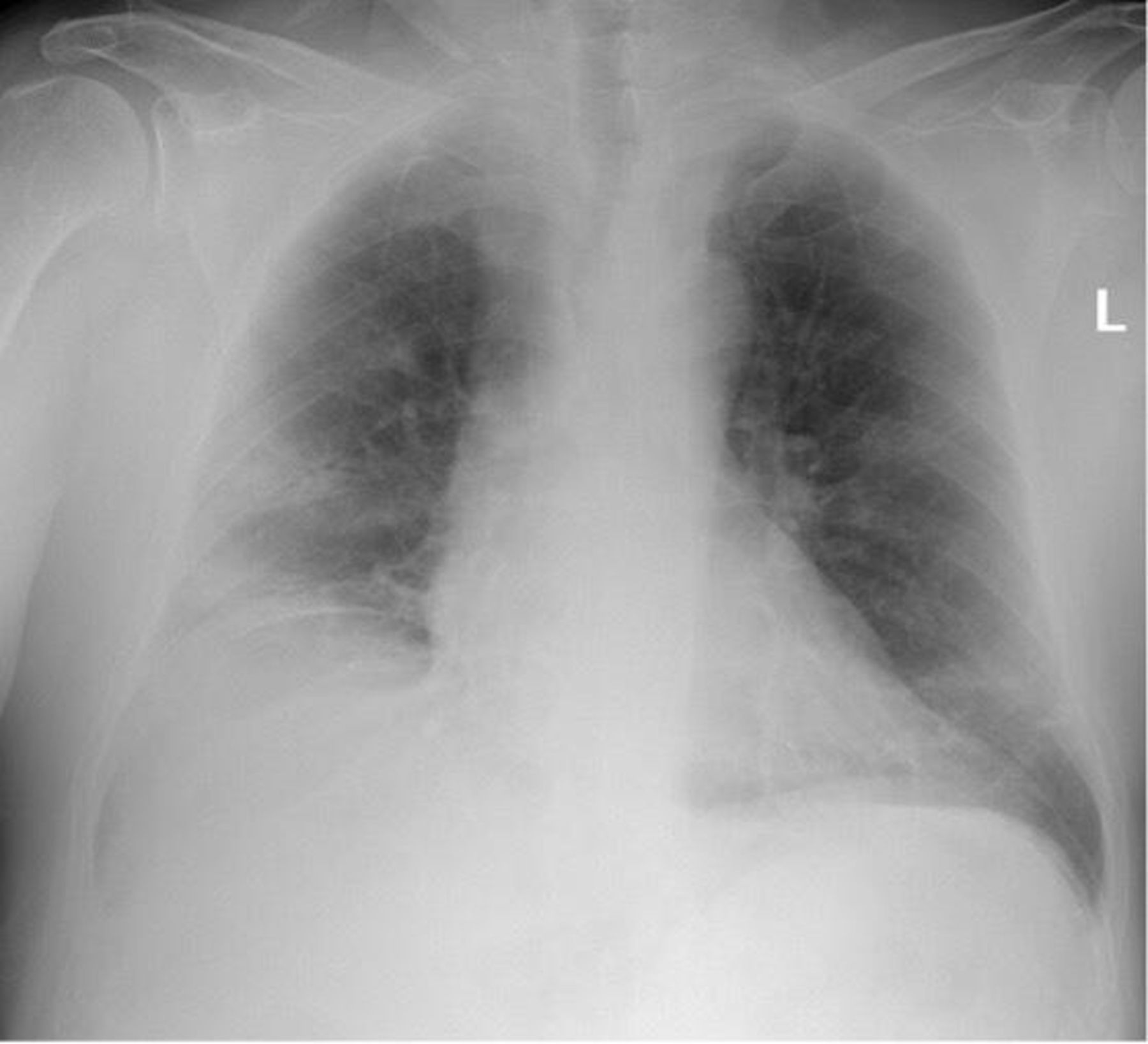
Chest radiography revealed peripheral cotton infiltrates in both lung bases with greater extension in the right base.
Treatment
According to the recommended protocol by our government’s health authority, Directorate-General of Health, the patient was started on oxygen and dexamethasone. Prophylactic enoxaparin, sodium chloride, furosemide, perindopril, paracetamol and metamizole were also administered.
Outcome and follow-up
In the following days, the patient promptly recovered, and we have been able to gradually reduce oxygen flow. On the day of discharge, the patient maintained the skin changes despite the resolution of COVID-19 symptoms. In the re-evaluation consultation, 30 days after discharge, complete resolution of the dermatological manifestations was noted.
Second case
Case presentation
A 57-year-old woman with a medical history of migraine, arterial hypertension, chronic venous insufficiency, gastro-oesophageal reflux disease and obesity, initially presented with diffuse urticarial rash on the cervical and chest region (figures 5 and 6). Three days later, she developed fever, fatigue and posterior thoracalgia.
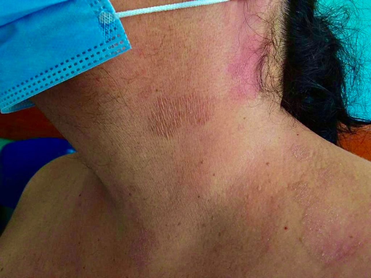
Urticarial rash on the cervical egion.
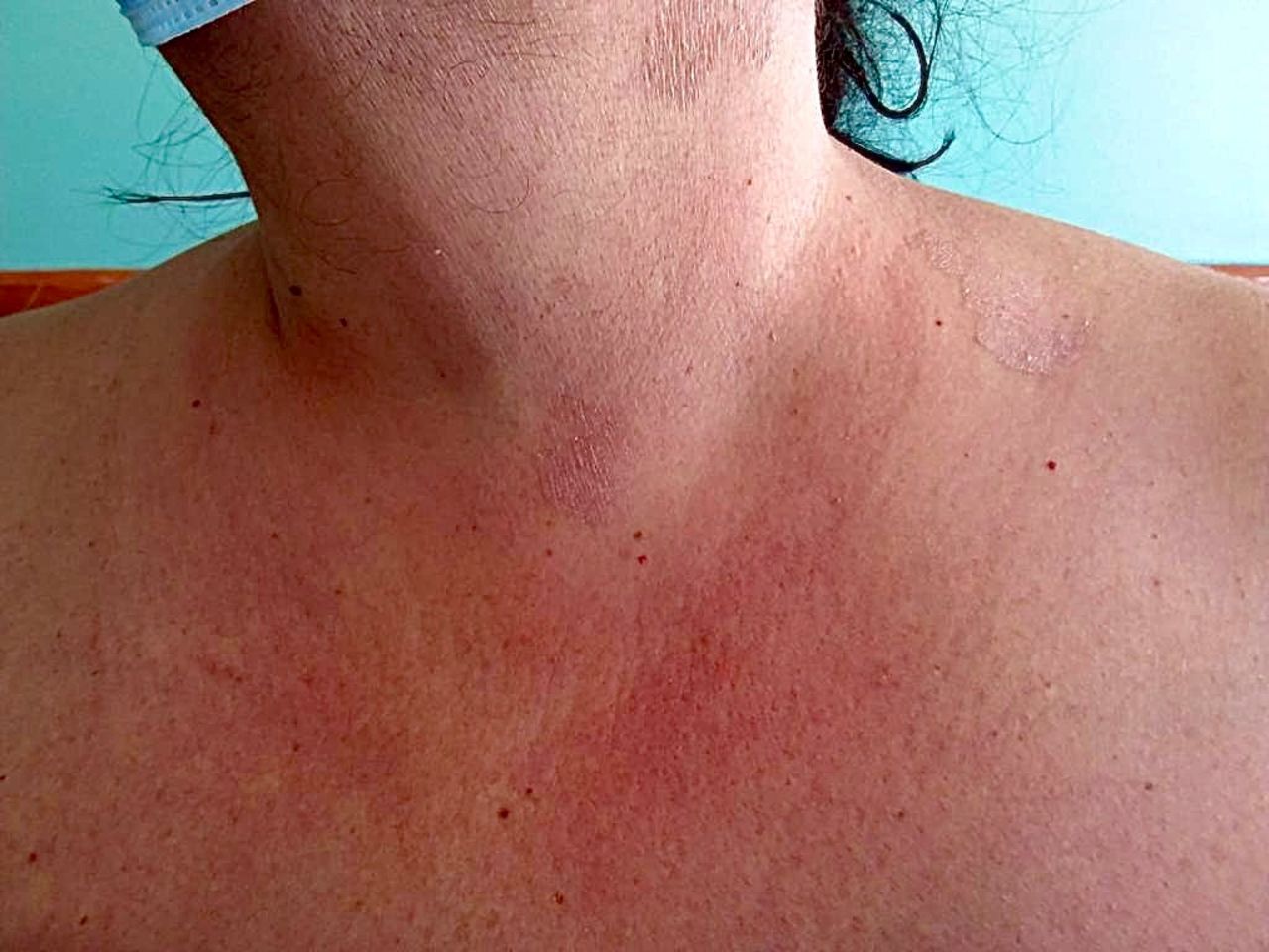
Urticarial rash on the cervical and chest region.
Investigations
Nasopharyngeal swab for SARS-CoV-2 RNA amplification came back positive on the day of admission to our COVID-19 ward. Blood test results revealed increased D-dimers (799 ng/mL), LDH (261 U/L), CRP (48.20 mg/dL) and ferritin (439 ng/mL). The white blood cells, platelet count, kidney and liver function, and ionogram were all normal. There were no signs of pneumonia on the chest X-ray.
Treatment
Oral cetirizine was started and the pruritus improved. Low-molecular-weight heparin in prophylactic dose (enoxaparin) and paracetamol were also administered.
Outcome and follow-up
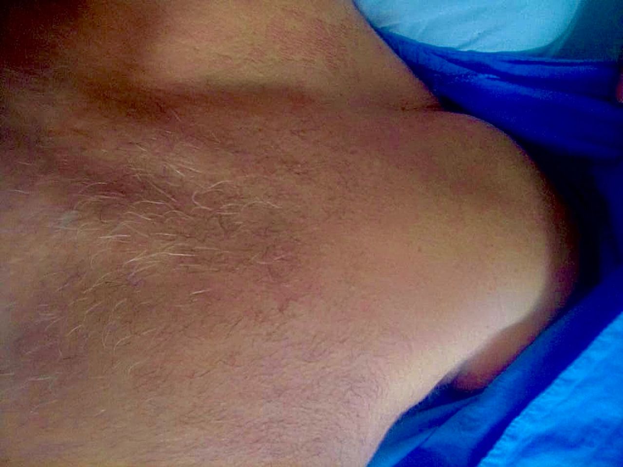
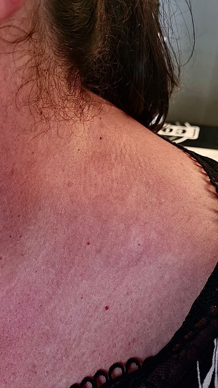
Thirty days after discharge, the patient maintained the rash, but without the pruritus (figures 7 and 8).

Urticarial rash 1 month after discharge.

Urticarial rash 1 month after discharge.
Third case
Case presentation
A 49-year-old man with a medical history of colon tubulovillous adenoma with high-grade dysplasia was admitted with thoracalgia, worsening shortness of breath and dry cough. He also had, on the cervical and chest region, urticarial rash (figure 9), which started 5 days before.
Urticarial rash on the cervical and chest region.
Investigations
SARS-CoV-2 infection, tested for by a nasopharyngeal swab, was diagnosed. Laboratory tests showed lymphopenia (700/µL), increased D-dimers (500 ng/mL), abnormal liver function (alanine aminotransferase 60.6 U/L, aspartate aminotransferase 115.9 U/L and GGT 108.4 U/L), elevated LDH (316 U/L), CRP (106.21 mg/L) and ferritin (2886 ng/mL). Platelet count, renal function and ionogram were normal. Chest X-ray was negative for pneumonia.
Treatment
The patient was treated with prophylactic enoxaparin, cetirizine and paracetamol.
Outcome and follow-up
The pruritus improved, and 5 days after discharge, the urticarial areas completely disappeared.
Discussion
The SARS-CoV-2 infection has caused numerous repercussions on millions of individuals worldwide, medically, financially and socially.2 Also, it constituted a new challenge to recognise the extensive range of clinical manifestations of the disease for healthcare professionals. Despite all the information currently released on this virus, much remains unknown about the disease’s clinical characteristics, including dermatological manifestations.
Articles from the beginning of the pandemic in Wuhan described the presence of rashes in 0.2% of 1099 confirmed cases of COVID-19,12 so they were considered of little relevance for several months.12 13 As the infection spread, reaching Europe, there was a vast recognition and interest in these category of manifestations.
The case series published by Recalcati in Lombardy1 was the first report of the cutaneous manifestations in COVID-19 patients during the severe acute respiratory syndrome. In this study, which included 88 patients, 20.4% (n=18) had skin lesions. The patterns described included erythematous (77.8%), urticarial (16.7%) and varicella-like eruptions (5.6%).1 12 After this publication, similar lesions were described in multiple clinical cases and case series, and other forms, such as petechiae lesions, perniosis-type, vasculitis, polymorphic erythema, livedo reticularis and reactivation of oral herpes simplex type 1 were also described.12 14 Although most of these rashes appear to emerge from the cytopathic effect of the virus, some cutaneous patterns may be the result of a deregulatory response by the immune and thrombogenic systems through the uncontrolled release of interferon and proinflammatory cytokines.2 12
Acral lesions, also defined as pseudochilblain, are described as a localised inflammatory dermatological disorder presented as erythematous–violaceous or purpuric macules on fingers, elbows, toes and the lateral aspect of the feet.6 These lesions have been observed to be more common in young adult patients with a milder disease course and manifesting after the onset of commonly COVID-19 symptoms.2 6 However, our patient from case 1, was older and, for the presence of bilateral pneumonia requiring oxygen therapy, SARS-CoV-2 infection was considered of moderate severity. Pseudochilblain eruptions are frequently asymmetrically distributed,15 and Galvan et al reported pain in 32% and itches in 30% of patients.6 Our patient had these lesions on both feet and reported only pain on palpation. Despite Singh et al stating an association between these lesions and exposure to cold temperatures or damp, humid environments,16 most studies did not confirm this relationship.2 7 12 17
The urticarial rash was the first dermatological pattern described in patients with SARS-CoV-2 infection12 and can appear before cough and fever.15 In the case series published by Recalcati, 16.7% of the 18 patients had these lesions, slightly itchy and mainly distributed over the trunk.1 12 It is important to note that these skin changes did not correlate with the severity of the disease12 and were described in patients with a milder clinical course and limited symptoms.13 In the literature cases, this rash has improved with the introduction of oral antihistamines.12 15 In our two patients with urticarial rashes, the COVID-19 had a milder course, and the introduction of cetirizine improved the associated pruritus.
Learning points
Acral areas of erythema–oedema with some vesicles or pustules (pseudochilblain), other vesicular eruptions, urticarial lesions, other maculopapules, and livedos or necrosis are the most reported cutaneous symptoms of COVID-19.
Clinicians should be aware of the cutaneous manifestations linked to COVID-19 as they might be the presenting sign of infection in asymptomatic or minimally symptomatic patients.
As in most of the aspects intrinsic to this new virus, much remains to be revealed concerning cutaneous manifestations, which demands further investigations.
Ethics statements
Patient consent for publication
Acknowledgments
The authors are grateful to our colleagues of COVID-19 ward, Sofia Gonçalves, Mariana Martins, João Paulo Correia, Miguel Santos, Pedro Balza, Pedro Mendes, and Ana Paula Reis, who collaborated in discussing the cases and were involved in drafting the manuscript.
Footnotes
Contributors HL carried out the conception of the manuscript, the acquisition of data on the patient in case 2, the literature search, the selection of information and the writing of the manuscript. CB carried out the literature search, the selection of information and helped in writing the manuscript. She also revised the manuscript. DA collected patient data from case 1 and 3. He helped in the literature search and the writing of the manuscript. AM carried out the literature search, analysis and interpretation of the collected data. She also contributed to the revision of the manuscript.
Funding The authors have not declared a specific grant for this research from any funding agency in the public, commercial or not-for-profit sectors.
Competing interests None declared.
Provenance and peer review Not commissioned; externally peer-reviewed.