Article Text

Statistics from Altmetric.com
Description
A 16‐year‐old boy with negative past medical history presented to an outside hospital with a first time right‐sided focal seizure. Neurologic examination at presentation was normal. CT performed in the emergency room revealed a large left intra‐axial cerebral mass without haemorrhage (figure 1A). Subsequent MRI demonstrated a large 7.5 cm well‐circumscribed intra‐axial mass within the left cerebral hemisphere with minimal enhancement and surrounding oedema (figure 1B–F). T2 shine‐through artefact on diffusion‐weighted imaging was initially misinterpreted as demonstrating reduced diffusivity, suggestive of hypercellularity, and high‐grade histology. The differential diagnosis based on the MRI features included high‐grade glioma, atypical teratoid/rhabdoid tumour, embryonal tumour, or anaplastic ependymoma. Atypical intra‐axial epidermoid tumour, complex neuroepithelial cyst, or low‐grade glioneuronal tumour were considered less likely diagnostic considerations. No fat density or calcifications were present on CT to suggest teratoma or dermoid cyst.

Neuroimaging features of epidermoid tumour. (A) Unenhanced CT demonstrates large irregular but well‐defined hypodense mass along the left Sylvian region (arrow) with mass effect and rightward midline shift. (B) Apparent diffusion coefficient map depicts predominantly atypical hyperintense increased diffusivity with only small central area of hypointense reduced diffusivity (arrow). (C) Unenhanced T1‐weighted MRI shows T1‐hypointensity (arrow). (D) T2‐weighted MRI demonstrates homogenous hyperintensity. (E) Fluid‐attenuated inversion recovery demonstrates heterogenous unusual hypointensity (arrow) without surrounding T2‐hyperintense oedema. (F) Post-contrast (gadolinium) T1‐weighted MRI reveals minimal peripheral linear enhancement, which likely reflects displaced adjacent vessels (arrow).
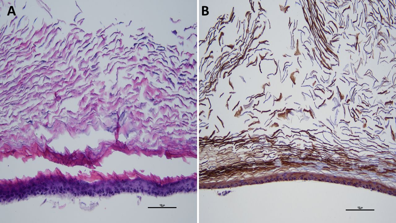
The patient underwent successful left craniotomy and gross total resection of large pearly white flaky tumour mass with thin translucent white membrane and areas of marginal mottled friable tissue. The membrane was adherent to the brain parenchyma. Neuropathology demonstrated no malignancy but abundant keratin with squamous epithelial lining that was positive for pan-cytokeratin by immunohistochemistry consistent with a diagnosis of epidermoid tumour (figure 2A–B). The patient suffered transient post‐operative aphasia that lasted 3 days with full recovery. There is no evidence of recurrent disease 12 months after diagnosis and he remains seizure free with a normal neurologic exam more than 1 year after diagnosis.
Neuropathologic features of epidermoid tumour. H&E stained section (A) demonstrates abundant keratin material with keratinised squamous epithelium along lining of cyst that was positive for pan-cytokeratin staining by immunohistochemistry (B) consistent with a diagnosis of epidermoid tumour.
Epidermoid cysts are benign complex cystic insinuating tumours which arise from abnormal ectodermal cell rests from embryogenesis. They grow slowly by desquamation of epithelial cells and accumulation of keratin and cholesterol.1 Less than 2% of intracranial tumours are reported as epidermoids which are encountered even less commonly in children. Most cases occur in the posterior fossa, and especially the cerebellopontine angle. Supratentorial intracerebral locations account for less than 1.5% of epidermoids but may be more common in children.1 2 Children most commonly present with headache but normal neurologic examination.2 On CT, epidermoids may appear identical to arachnoid cysts. On MRI, they characteristically exhibit isointense or mildly hyperintense signal on T1‐weighted images, and hyperintensity on T2 and fluid‐ attenuated inversion recovery (FLAIR) sequences. Reduced diffusivity on diffusion-weighted imaging (DWI) is regarded as pathognomonic due to restriction of free motion of water molecules by the concentric keratin layers.3 Interestingly, our case does not exhibit this specific MRI feature unlike all reported in the only series exclusively limited to paediatric intracranial epidermoids.2
Complete neurosurgical resection can be challenging with postoperative risks of stroke, cranial neuropathies, chemical meningitis (due spillage of cyst contents), hydrocephalus, and 24% risk of recurrence.4 5 Less than 50 cases of extremely rare malignant degeneration of intracranial epidermoids into squamous cell carcinoma have been reported with only one in a child.6 Our case highlights the diversity of MRI findings associated with intracranial epidermoid tumours.
Learning points
Epidermoid tumours are benign neoplasms rarely seen in children. They are typically found as extra-axial posterior fossa masses with reduced diffusivity on diffusion weighted imaging and apparent diffusion coefficient map, and hyperintensity on fluid‐attenuated inversion recovery (FLAIR) sequence on MRI.
We report a highly unusual paediatric case of epidermoid presenting as a large intracerebral mass with significant mass effect, and very atypical increased diffusivity and FLAIR-hypointensity on MRI.
Lack of surrounding vasogenic oedema despite significant mass effect, and hypodensity of mass on CT should encourage inclusion of epidermoid as a differential consideration despite atypical intra-axial location and atypical MRI features.
Ethics statements
Footnotes
Contributors EGC was responsible for the design and writing of the manuscript. DM was responsible for the design and writing of the manuscript. ML was responsible for the design and writing of the manuscript. JRC was responsible for the design and writing of the manuscript.
Funding The authors have not declared a specific grant for this research from any funding agency in the public, commercial or not-for-profit sectors.
Competing interests None declared.
Provenance and peer review Not commissioned; externally peer reviewed.