Article Text

Statistics from Altmetric.com
Description
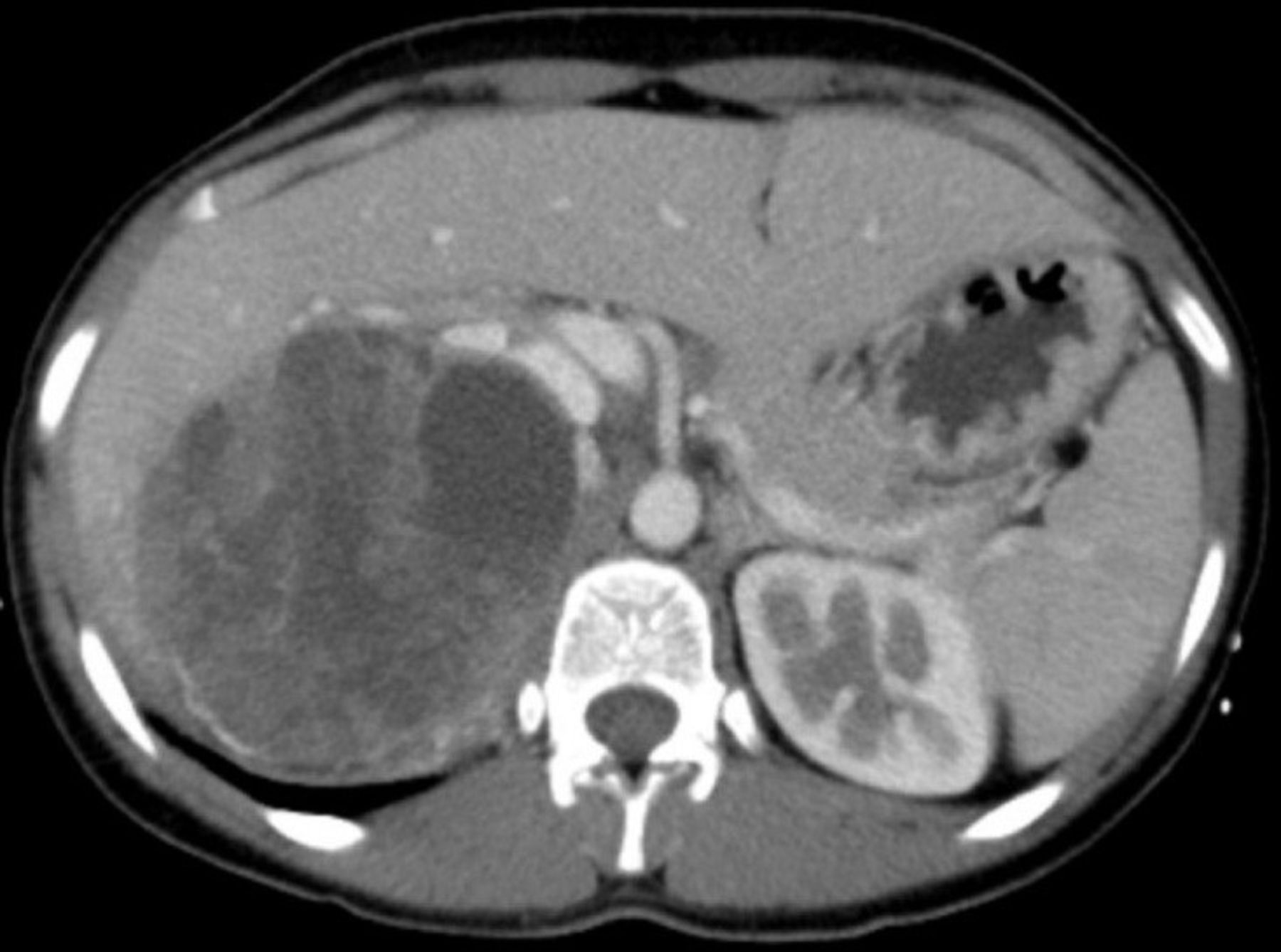
A 22-year-old woman presented to the urology clinic with a 6-month history of right flank pain with decreased appetite, nausea, progressive fatigue and unintentional weight loss. Surgical history was significant for the right simple renal cyst decortication 10 years ago. Medical history was otherwise not significant. Physical examination revealed a right flank mass with mild tenderness, in addition to the right flank surgical scar. Laboratory tests were notable for microscopic haematuria (60–70 RBCs/field). Urine cytology did not reveal malignant cells. Renal ultrasound showed a right kidney hyperechoic mass with a cystic solid appearance. A multislice CT confirmed the presence of a complex cystic solid mass without contrast enhancement compatible with type IV of Bosniak classification. The mass was infiltrating the right haemidiaphragm and liver and compressing the inferior vena cava (figures 1 and 2). The patient underwent right radical nephrectomy through a right subcostal transperitoneal approach. Macroscopically, a well-demarcated solid mass was located in the mid and upper portions of the kidney and measured 12.5 cm in addition to numerous cysts with various sizes containing clear fluid, resulting in a spongy-like appearance (figure 3). Pathology confirmed the diagnosis of tubulocystic renal cell carcinoma (TC-RCC) (figure 4) along with areas of papillary RCC (figure 5). Immunohistochemical profile of the pathology specimen was positive for CD10, weakly positive for pancytokeratin and vimentin and negative for CK7 and CK20, further confirming the diagnosis of RCC.

Coronal view of CT abdomen and pelvis without contrast enhancement at the level of upper pole of the right kidney revealing the solid part of mass with a cystic formation. Note the mass compressing the inferior vena cava and infiltrating the hepatic tissue.

Coronal view of CT abdomen and pelvis without contrast enhancement at the level of mid-pole showing several cystic formations.

Gross view of the right kidney shows well-demarcated mass involving the upper two thirds of the kidney. The cut surface shows spongy tan tissue with multiple cysts in various sizes and partial haemorrhage and infarctions.

Microscopic examination of TC-RCC using high power view shows tubules or cystic structures lined by a single layer of epithelial neoplastic cells with eosinophilic cytoplasm. TC-RCC, tubulocystic renal cell carcinoma.

Microscopic view of the papillary RCC combined component. RCC, renal cell carcinoma.
TC-RCC is a rare form of RCC with <100 cases reported in the literature.1 According to the 2016 WHO classification, TC-RCC is an independent class of RCC. TC-RCC affects predominantly men in their fifth and sixth decades with male to female ratio of 7:1. Although patients with TC-RCC might present with abdominal pain, distention, haematuria and weight loss, the vast majority is asymptomatic. Clinically, behaviour of TC-RCC is typically indolent. However, aggressive tumours were reported in about 10% of cases. Gross pathology of TC-RCC reveals well-circumscribed multicystic lesions that are frequently illustrated as ‘bubble wrap-like’ or ‘sponge-like’ with no capsule. Microscopically, TC-RCC comprises tubules and cysts of variable sizes, which are lined with a single layer of cells with eosinophilic cytoplasm and show intervening septa. Interestingly, the coexistence of TC-RCC with papillary RCC is not uncommon.1 2 The coexistence of poorly differentiated foci suggests worse prognosis.3 Differential diagnoses for TCC include cystic nephroma, hereditary leiomyomatosis and RCC, multilocular cystic renal cell carcinoma, mucinous tubular and spindle cell neoplasm. Radical or partial nephrectomy depending on the tumour size and location are considered the gold standard of management. The prognosis is usually excellent with rare recurrence and low mortality.1
Learning points
Tubulocystic renal cell carcinoma (TC-RCC) is a rare subtype of RCC with indolent symptoms and excellent prognosis.
The association of TC-RCC with papillary RCC is not uncommon.
Being familiar with the macroscopic and microscopic features of TC-RCC is considered very helpful in making the diagnosis and determining prognosis.
Footnotes
Contributors AhA, AA and AD: conception and design of this report, acquisition of data as well as drafting the manuscript. AI: contributions include drafting the report as well as critically revising it. All co-authors approve this manuscript for final publication. All authors certify that they have participated sufficiently in the work to take public responsibility for the content, including participation in the concept, design, analysis, writing or revision of the manuscript.
Funding The authors have not declared a specific grant for this research from any funding agency in the public, commercial or not-for-profit sectors.
Competing interests None declared.
Provenance and peer review Not commissioned; externally peer reviewed.
Patient consent for publication Obtained.